 Pancreas
-- a large exocrine gland for secretion of digestive enzymes that
are carried by a duct system to the duodenum. Small areas of
endocrine tissue, the islets of Langerhans, will be studied later. Pancreas
-- a large exocrine gland for secretion of digestive enzymes that
are carried by a duct system to the duodenum. Small areas of
endocrine tissue, the islets of Langerhans, will be studied later.
Examine sections of pancreas (slide
15,
44 and
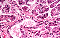
154) with low power and observe the overall organization into
lobules separated by CT septa with adipocytes, the pale-staining
pancreatic islets (of Langerhans) surrounded by densely packed acini, and the
excretory ducts (Fig. 16-8).
What are the cells that
comprise most of each lobule called?
How do pancreatic acini and
pancreatic islets differ structurally and functionally?
How are the cells of pancreatic
ducts different and how are they similar to the same cells in
salivary glands?
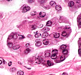 With
the 40X objective, examine acini in an area where the polarized
nature of the cells is apparent (Fig. 16-9) With
the 40X objective, examine acini in an area where the polarized
nature of the cells is apparent (Fig. 16-9)
- Note that the lumens of
pancreatic acini are almost too small to see with the light
microscope. Compare the appearance of acini with the electron
micrograph in Fig. 16-10.
How does the pancreas
neutralize the acidic chyme entering the duodenum from the stomach?
Next is the
liver. |