| Describe the formation of the
ovarian
structures, listed on the previous page, in process of follicular development, i.e., how a
mature follicle is formed.
Sketch an oocyte and the cumulus oophorus in a mature follicle and
label all the structures.
Clincial note: Menopause
results when the supply of ovarian follicles is exhausted at about
age 50. With loss of the steroid hormones secreted by the follicles,
cyclic growth and loss of the uterine endometrium stops and a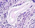 variety of somatic and emotional problems can occur.
On the same slide, identify atretic
follicles which stopped developing at various stages (Fig. 22-9).
What is the significance of
follicular atresia?
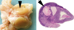 Identify
the corpus luteum on slide 13 and the very large corpus luteum on
slide 58 (Fig.
22-10). Within the corpus luteum on
slide 58, identify
the masses of granulosa lutein cells, separated partially by septa,
and surrounded by a thinner layer of less prominent theca lutein
cells (Fig. 22-10). Identify
the corpus luteum on slide 13 and the very large corpus luteum on
slide 58 (Fig.
22-10). Within the corpus luteum on
slide 58, identify
the masses of granulosa lutein cells, separated partially by septa,
and surrounded by a thinner layer of less prominent theca lutein
cells (Fig. 22-10).
Based on their histological
appearance, what do these cells secrete? What is their function?
Examine
slide 61 and identify a
corpus albicans (Fig. 22-11). This ovary specimen from an older
individual contains few follicles, but several corpora albicans.
What does the corpus albicans
represent and why do these structures persist much longer in
mothers?
Now for the
oviducts.
|