Cardiac Muscle
Fibers of cardiac muscle have features
somewhat intermediate between those of skeletal and visceral muscle.
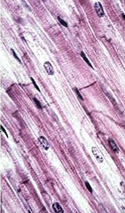
- Examine muscle in the heart
myocardium (slide 64 and
65).
- Identify fascicles, connective
tissue layers, blood microvessels, and fibers cut in both
directions (Figs. 10-16 through 10-18).
- Be sure to identify intercalated
disks and distinguish these from striations.
Study electron micrographs of cardiac
muscle (Figs. 10-17 and 10-18).
- Note the similarities and
differences with skeletal muscle, particularly the intercalated
disks.
- Note also the secretory granules
with atrial natriuretic factor that are present in some atrial
myocytes.
- Compare and contrast the
structural features of cardiac muscle with the other two muscle
types and indicate the functional significance of these
features.
 Clinical
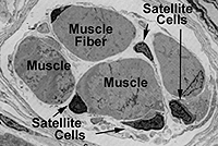
note: The fact that cardiac muscle lacks satellite cells
severely limits its capacity for repair after injury, such as injury
from transient ischemia during myocardial infarction (heart attack).
As shown on the next page, injured cardiac myofibers degenerate and
are replaced by connective tissue. Cardiac muscle injury and
degeneration causes release of various cytoplasmic enzymes, which
can be detected in the circulating blood and used as an indicator of
heart damage. Clinical
note: The fact that cardiac muscle lacks satellite cells
severely limits its capacity for repair after injury, such as injury
from transient ischemia during myocardial infarction (heart attack).
As shown on the next page, injured cardiac myofibers degenerate and
are replaced by connective tissue. Cardiac muscle injury and
degeneration causes release of various cytoplasmic enzymes, which
can be detected in the circulating blood and used as an indicator of
heart damage.
Sketch the cells/fibers of the
three types of muscle to the same scale.
Now on to
nervous tissue. |