Mucosa-associated Lymphoid Tissue
(MALT)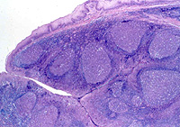
Examine a section of tonsil (slide 136,
Fig. 14-15). Identify the stratified squamous epithelium covering
the tonsil and the dense connective tissue at its base. Identify the
large lymphoid follicles. The large, pale cells scattered among the
lymphocytes in the follicles are macrophages acting as
antigen-presenting cells.
Why might you expect there to
lots of APCs in tonsils?
 Clinical
note: Tonsillitis, pharyngitis, and other upper respiratory
tract infections are among the most common problems involving the
head and neck. Tonsillectomies, which were formerly much more common
than now, may be required if chronic tonsillitis causes enlargement
of the affected structures to the degree that air passages are
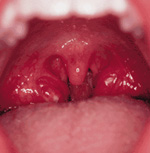
obstructed. To the right we see a pair of very large and inflamed
tonsils. Clinical
note: Tonsillitis, pharyngitis, and other upper respiratory
tract infections are among the most common problems involving the
head and neck. Tonsillectomies, which were formerly much more common
than now, may be required if chronic tonsillitis causes enlargement
of the affected structures to the degree that air passages are
obstructed. To the right we see a pair of very large and inflamed
tonsils.
Examine the connective tissue in the
wall of the ileum (slides 27 and
37) and identify the nodule of
lymphoid tissue representing a Peyer's patch (Fig. 14-16). Similar
diffuse nodules of various sizes will be seen in sections of
esophagus and many regions of the small and large intestines.
What is one difference between
the lymphoid tissue of tonsils and Peyer’s patches?
Identify the nodules of lymphoid
tissue in the wall of the appendix (slide 41, Fig.
15-39).
What are some distinguishing
features of the appendix?
What is the general purpose of
MALT along the gut?
The spleen is
next. |