Thick skin -- present on
fingertips, palms, and soles of feet, it is specialized for
protection during friction and contains sensory receptors responsive
to pressure, the Pacinian corpuscles.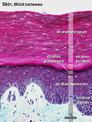
Examine a section of thick skin (slides
5 and
89) and identify epidermis and
dermis (Figs. 18-1 through 18-3).
- In the dermis, note the difference
between the papillary and reticular layers.
- Note too how downward epidermal folds or
ridges interdigitate with upward projecting dermal papillae.
- In the epidermis, identify
characteristic cells, keratinocytes, (as seen in Fig. 18-2)
representing the
- Stratum basale,
- Stratum spinosum,
- Stratum granulosum, and
- Stratum corneum
- Click image to right for
expanded view.
- The strata grade into one another
and cells gradually move outward from the basal layer to the
cornified layer.
What is the structural
significance of the small “spines” on the cells of the spinous
layer?
Study the ultrastructural features of
the intercellular connections of the keratinocytes (Fig. 18-4).
Describe the cell biology of
the keratinization process, accounting for all the histological
features seen.
The skin as a route of
medication delivery. |