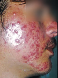
 Clinical
note: Sebaceous glands become much more active after puberty and
can become affected by acne vulgaris, in which blocked ducts or hair
follicles produce small white lesions (comedones) caused by build-up
of sebum and dead cells. The disease is normally self-limiting, but
comedones can become sites of bacterial infections. Sometimes there
is marked scaring left in the wake of the inflammation. Clinical
note: Sebaceous glands become much more active after puberty and
can become affected by acne vulgaris, in which blocked ducts or hair
follicles produce small white lesions (comedones) caused by build-up
of sebum and dead cells. The disease is normally self-limiting, but
comedones can become sites of bacterial infections. Sometimes there
is marked scaring left in the wake of the inflammation.
On slides
36 and
89 identify the
- Secretory portions and
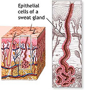
darker-staining ducts of merocrine (“eccrine”) sweat glands (Figs.
18-16a and 18-17).
- Myoepithelial cells surrounding
the secretory units (better shown on
slide 89).
- In the ducts, note the unusual
stratified cuboidal epithelium lining the ducts and the lack of
myoepithelial cells.

Examine the micrograph of apocrine
sweat glands (Fig. 18-16b) and look for such structures on
slide 17 and
slide 25.
- Compare the lumen size and overall
histological appearance of apocrine sweat glands to the more
common merocrine/eccrine sweat glands.
Summarize the histological and
functional differences between merocrine and apocrine sweat glands.
Examine the structure of nails and
their association with other parts of a digit as shown on the demo
slide (Fig. 18-14).
How does the structure of a
nail and nail bed compare with that of a hair follicle?
Next is the
cardiovascular system. |