Take a look at several slides and
describe what you see.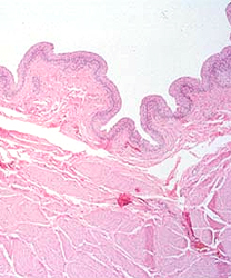
- Examine any three
different slides stained with H&E and identify basophilic
and acidophilic structures.
- Then choose two similar tissues
stained individually with H&E and with Masson's trichrome,
- For example, slides
28 and
12 of bladder.
- Examine the slides under low
power and compare the staining properties shown by the two
methods.
- You should start to become
familiar with the properties associated with these common stains
and be able to recognize the most common staining techniques
used on your slides.
What are the major colors you
see in slides stained with H&E?
What are the major colors you
see in slides stained with Masson’s trichrome?
What are the relative advantages of each of these two staining
methods?
Interpretation of a slide requires
a
systematic approach. |