|
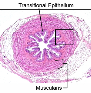 |
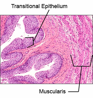
The ureters have walls with three principal layers. Use
this virtual microscopic slide of URETER to identify the ureter and the
following components:
- The inner mucosa, including a surface of transitional
epithelium and underlying areolar connective tissue.
- Muscularis, containing smooth muscle, with inner layers
arranged more longitudinally and outer layers more circularly.
- The outer adventitia, which is formed from areolar
connective tissue.
|