|
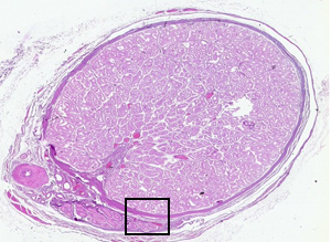 |
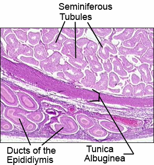
In this TESTIS virtual microscopic slide note the thick tunica albuginea of dense connective tissue surrounding the
testis.
Much thinner connective tissue septa divide the testis into
lobules.
- Seminiferous tubules are found within each lobule. Under
higher magnification, the cells of these tubules can be seen at
the various stages of spermatogenesis.
- Spermatogonia are located along the basement membrane
of each tubule.
- Sustentacular cells have a pale-staining, often
triangular nucleus with a small darker nucleolus.
- Interstitial cells (of Leydig) are found in groups within
the connective tissue which surrounds the seminiferous tubules.
The interstitial cells produce the hormone testosterone and thus
are the endocrine portion of the testes.
- The little box indicates where the picture below came
from.
|