Anatomy A215 Virtual
Microscopy
|
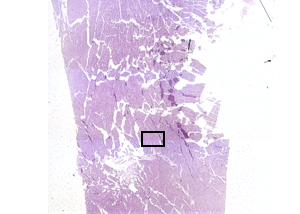 |
This virtual microscope slide is of a piece of human HEART. It
includes not only the muscle of the heart, but also the outer
covering known as the epicardium. The little box in the image to
the left indicates where the photograph below came from. This is in
the middle of the cardiac muscle and would be a good place to find the
features you need to be able to identify. |
|
 |
Cardiac muscle cells have a
cylindrical shape and may branch. Each has one, or possibly two,
nuclei along the axis of the cylinder. Cross striations,
perpendicular to the axis, are present. Cells meet end-to-end at
specialized junctions (gap junctions) called intercalated discs
which are parallel to, but darker than, the faint striations.
|
A215 Home Page
| Virtual Microscopy
Table of Contents
|