Anatomy A215 Virtual Microscopy
Skeletal Muscle - Longitudinal Section


This SKELETAL MUSCLE - LONGITUDINAL SECTION slide shows the large diameter cylindrical cells referred to as skeletal muscle fibers Each has many nuclei scattered along its length and located immediately under its cell membrane.

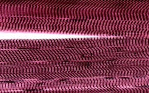
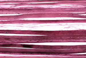
Internally, a muscle fiber has lengthwise myofibrils, each composed of a series of segments called sarcomeres. Each sarcomere contains many lengthwise myofilaments. The two types of myofilaments (thick & thin) occur in a regular arrangement which causes the cross striations (alternating dark and light bands) to be seen in myofibrils and fibers. These bands are readily seen in the image to the left.

See this slide with the virtual microscope

I don't see the virtual slide, I must need the download.


   A215 Home Page | Virtual Microscopy Table of Contents