|
 |
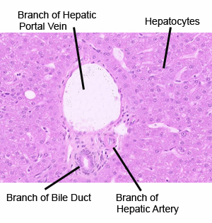
Each lobe of the liver is divided into many lobules, each
formed by radially arranged cords or plates of hepatocytes (liver
cells). In this LIVER virtual slide, identify the
following:
- Lobule, roughly hexagonal in cross section.
- Central vein, in the center of a lobule.
- Hepatic triad (at each of a lobule’s corners) and its three
components:
- A branch of the hepatic portal vein.
- A branch of the hepatic artery (often an arteriole).
- A branch of the bile duct, lined by simple cuboidal
epithelium.
- Sinusoids, thin-walled vascular spaces draining toward
the central vein.
- Hepatocytes, the main cell type in the liver.
|