 Esophagus
-- conducts food from oral cavity to stomach. Start by reviewing the
general plan of the gastrointestinal tract (Fig. 15-2), noting
especially the four major layers which are clearly seen in the
esophagus: Esophagus
-- conducts food from oral cavity to stomach. Start by reviewing the
general plan of the gastrointestinal tract (Fig. 15-2), noting
especially the four major layers which are clearly seen in the
esophagus:
- Mucosa
- The epithelial lining of various
types
- Lamina propria of connective
tissue
- Thin muscularis mucosae of
smooth muscle
- Submucosa containing blood
vessels, lymphatics, and nerves
- Muscularis: two thick layers of
smooth muscle for peristalsis
- Adventitia or serosa: outer
connective tissue covering
Examine the preserved-mounted
specimens of the various regions of the digestive tract, noting the
macroscopic features (stomach rugae, plicae circulares, etc.) for
correlation with the slides you will also study.
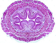
Examine a transverse section of the
esophagus. Identify the various
layers and sublayers indicated in the general plan.
- Note the epithelial type of the
lining,
- The presence of small mucous
glands
- Any lymphoid nodules present, and
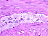
- Autonomic ganglia and nerves of
the myenteric plexus.
What type of muscle do you see in slides of esophagus and what does
this tell you about the level from which the biopsy was taken?
Let's consider the
muscular layers in more detail. |