
General
and Systemic Histopathology, C601&C602
Slide 53: Dissecting aortic aneurysm
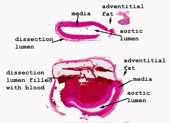 |
It may be a little hard
to imagine what's happening here, but if you study this scan of the tissue
and try to sketch, it may become clear. Somewhere in the wall of the aorta,
a tear in the intima developed (no, you can see the tear in this picture),
allowing the blood to "dissect" into and separate the muscle layers of
the aortic wall. Here we see what appears to be a "double barreled" lumen,
but in fact only one "true" aortic lumen is present. The other blood
filled space is the compartment created by the tearing and separation of
the aortic wall by means of the blood pressure pushing the blood into the
wall through the intimal defect.
See this slide with the
virtual microscope.
|
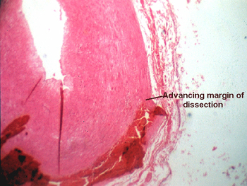 |
Again, look at this slide
on a white background first. You should easily be able to see the advancing
edge of the dissecting aneurysm. This condition may complicate an atherosclerotic
aneurysm, but may also be seen in the absence of atherosclerosis. It sometimes
happens in people with hypertension. Typically it is very painful as the
leading edge of the dissection tears the muscle of the aorta. The dissection
may stop, "blow" through the wall into the surrounding fat or pericardial
space, or even open another "rent" in the intima and reenter the lumen
of the aorta. |
Back
to Home
|