General
and Systemic Histopathology, C601&C602
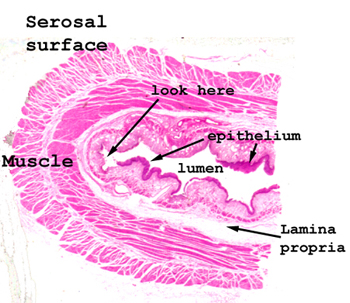
Slide 19: Acute Erosive Esophagitis
 |
Your slide shows almost
a complete cross section of the esophagus. Note there are several
areas of mucosal ulceration and some relatively large, thin walled vessels
are present in the lamina propria.
See this slide with the
virtual microscope. |
 |
If you simply look at
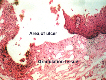
the slide on a white background, you can see the area of erosion quite
nicely. Note the areas of healing and repair at the margins of the ulcer.
Some acute inflammatory cells are seen in the base of the lesion, and many
very dilated vessels are present in this area as well. These large superficial
vessels are not part of the healing process, and represent part of another
pathologic process in this person. What condition
could lead to these dilated vessels? Where would you expect to find
others? |
Back
to Home
|