
General
and Systemic Histopathology, C601&C602
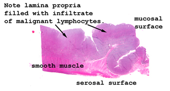
Slide 167: Intestinal lymphoma
|
Again, look how obvious
the lesion appears in this picture. You can easily see the lymphoid
infiltrate and muscular wall. It may be that your slide doesn't include
any "normal" bowel. If that's the case, I'm sorry, look at one of the
two ends of the section and I think you'll see something approximating normality.
See this slide with the
virtual microscope.
|
|
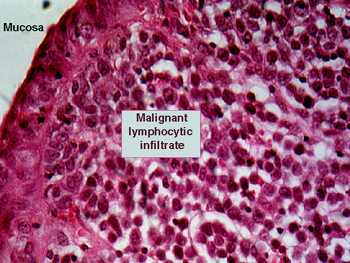
This is the second
such example of this type of tumor in your set. The features are essentially
the same as in slide 142. See which is the better example in your set and
spend time with that one. You will see the malignant lymphoid infiltrate in
the submucosa and possibly extending through the muscular wall. Note the
monomorphic nature of the malignant lymphocytes. The distinction of nodular
and diffuse does not apply here; these terms only have reference in lymph
nodes themselves. |
Back to Home
|