
General
and Systemic Histopathology, C601&C602
Slide 43: Heart with Myocardial Infarction
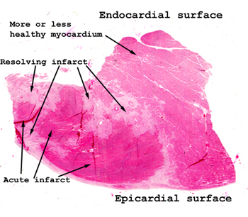 |
There's a lot to be seen
in this piece of tissue. We have more or less normal heart tissue right
beneath the endocardium. There are several myocardial infarctions
of various ages and demonstrating various stages of development. In this
picture, the areas identified as "resolving infarct" consist mostly of
granulation tissue and represent an infarction of about two to three weeks
duration. The bright pink areas represent a second and much more
recent infarction. After you've read the section on myocardial infarctions,
see if you can establish the duration of this second one. What do
you think was the most likely cause of death?
See this slide with the
virtual microscope. |
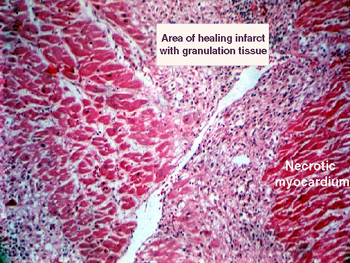 |
By this time it should
be obvious that a lot can be learned from a slide without using the microscope.
See if you can find the area of infarction before putting the slide on
the stage of your microscope. Hold it to the light or put it on a white
background. This area of infarction is about two weeks old, and shows substantial
removal of the dead muscle with early replacement with granulation and
fibro-connective tissue. Some inflammatory cells remain. This person died
with an arrhythmia. |
Back
to Home
|