
General
and Systemic Histopathology, C601&C602
Slide 59: Colon with acute ulcerative
colitis
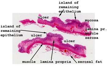 |
This can be a challenging
slide. There are only islands of remaining mucosa, separated by large expanses
of ulceration. Look in the base of the ulcers and right at the edge
of the residual mucosa for the best examples.
See this slide with the
virtual microscope. |
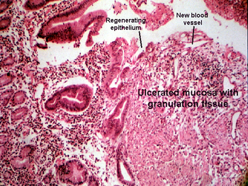 |
This is a higher power
view of the previous slide. It highlights the areas of neovascularity.
Pretty much everything applies here as was said about the previous one.
Here you can see the newly developing blood vessels a little better, but
still the intensity of the mixed inflammatory infiltrate may be hard to
read through. |
Back
to Home
|