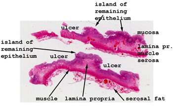
This can be a challenging slide. There are only islands
of remaining mucosa, separated by large expanses of ulceration. Look
in the base of the ulcers and just to the edge of the residual mucosa for
the best examples of granulation tissue.
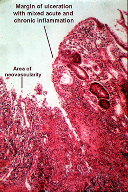
The picture is from the edge of an ulcer, and shows
focal absence of the mucosa. There is a mixed acute and chronic inflammatory
infiltrate in the base of the ulcer with lots of granulation tissue. You
will see many angioblasts and reactive fibroblasts in these areas of healing.
Crypt abscesses are seen in the base of the crypts. This feature may not
be very apparent as this case is relatively advanced and these earlier
changes are simply overwhelmed by the degree of inflammation.