General
and Systemic Histopathology, C601&C602
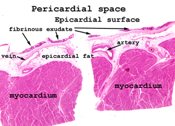
Slide 48: Fibrinous pericarditis
 |
In this case, we're looking
for a thin band of homogeneous, pink staining, proteinaceous material on
the epicardial surface of the heart. This represents an exudate composed
largely of protein material. You will see very few inflammatory cells.
See this slide with the
virtual microscope. |
 |
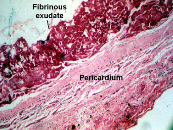
Now you can see how devoid
of inflammatory cells this exudate is. Remember it consists almost totally
of protein (fibrin plus other trash). It looks the way I think "tofu" would
look if sectioned and stained. |
Back
to Home
|