
General
and Systemic Histopathology, C601&C602
Slide 1: Cryptococcal Pneumonia
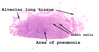 |
This is a picture of
the tissue as it appears on your slide. See if you can orient it
as it appears here and then locate the alveolar lung tissue with central
area of inflammatory infiltrate. You might even be able to spot some
of the giant cells without any magnification. When you put the slide
on the stage of your scope, start with the lowest power first, review the
entire slide and then go progressively to the higher levels of magnification.
See this slide with the
virtual microscope. |
 |
In this medium power
magnification, one can observe the quality and major constituents of the
inflammatory infiltrate. You will see it consists almost exclusively of
lymphocytes, plasma cells and monocytes. It is often referred to as a "mononuclear
infiltrate" because the cells that comprising it do not have lobated nuclei.
This slide depicts a process in which a special variety of inflammatory
cell is called into existence, the "giant cell." Because of the presence
of the giant cells, this pattern is also referred to as "granulomatous
infiltrate." Well developed granulomas are not seen in this case.
You can, however, see the giant cells containing organisms. |
Back
to Home
|