
General
and Systemic Histopathology, C601&C602
Slide 1: Cryptococcal Pneumonia, a
higher power view.
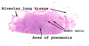 |
This is a picture of
the tissue as it appears on your slide. See if you can orient it
as it appears here and then locate the alveolar lung tissue with central
area of inflammatory infiltrate. You might even be able to spot some
of the giant cells without any magnification. When you put the slide
on the stage of your scope, start with the lowest power first, review the
entire slide and then go progressively to the higher levels of magnification.
See this slide with the
virtual microscope. |
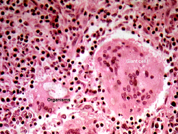 |
This is a second picture
form the same slide. As you can see the term "giant cell" is aptly applied.
The cryptococcal organisms are quite evident in one of the smaller giant
cells. As you may recall, the organisms posses a large capsule so they
tend to stand out in the cytoplasm of the giant cells. The giant cells
are unique participants in our response to this type of injury. They
are commonly seen in association with agents the body cannot easily rid
itself of. We will see them again in the inflammatory response to tuberculosis
and foreign material that has been injected or left behind in the body.
Can you think of situations in which foreign material might find its way
into the body (I mean external matter, not an infectious agent)? Here
are a few. |
Back
to Home
|