
General
and Systemic Histopathology, C601&C602
Slide 29: Hepatic adenoma
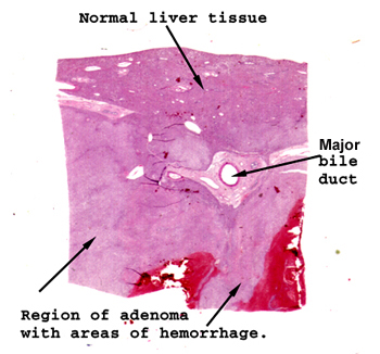 |
It's a little tricky
to see the area of the adenoma if all you do is slap the slide on the stage
of your scope. See if you can match the areas of the tissue as depicted
to the left and then look with your microscope right at the margin of the
tumor. With situations like this, it's really important to see both
cell types in one field.
See this slide with the
virtual microscope.
|
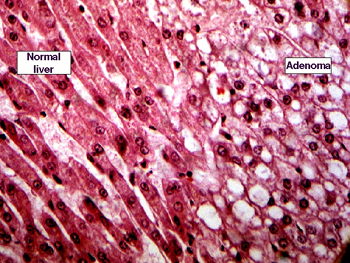 |
Here you can see the
cellular differences a little more clearly. The cells that make up this
benign tumor do not show much of a lobular arrangement. There are no triads.
The individual cells have a "foamier" cytoplasm and somewhat more vesiculated
nucleus. I don't think you will find any mitosis. These can become symptomatic
by bleeding, and can even cause death by this mechanism. What is associated
with these? Hint: think common exogenous hormones women may take. |
Back
to Home
|