
General
and Systemic Histopathology, C601&C602
Slide 103: Adenocarcinoma of gallbladder
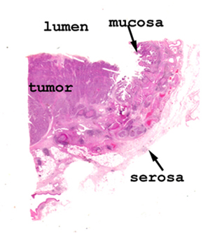 |
Not much trouble seeing
the cancer here. When you look at the tissue on the slide, you should
be able to see the uninvolved mucosa, muscular wall and serosal surface.
Use that area to get oriented and then move to the area of the adenocarcinoma.
See this slide with the
virtual microscope. |
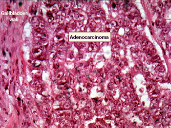 |
I don't think there is
much in the way of normal gallbladder left on this slide. It might help
if you look around on your slide to find some to get oriented. The malignant
cells of the neoplasm are very undifferentiated and are not forming much
in the way of glands. Seeing the hallmark cytologic features of malignancy
will be very easy. What are they? Predictable consequences of this condition? |
Back
to Home
|