
General
and Systemic Histopathology, C601&C602
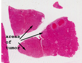
Slide 63: Liver with hepatocellular carcinoma
|
I think it is possible
to see the nodules of malignancy even with no magnification. If nothing
else, you can see areas of the liver tissue are distinctly different from
one another.
See this slide with the
virtual microscope.
|
|
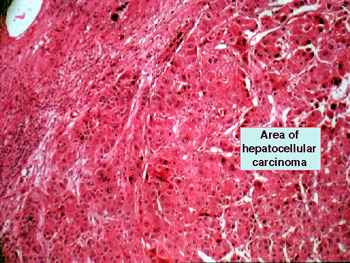
This cancer generally
arises in a background of cirrhosis and represents a primary malignancy
of the hepatocyte. You will see there is no lobular organization in the area
of the tumor. Note the marked degree of nuclear atypia and the great number
of mitoses. You are also likely to see many bizarre mitotic figures. There
may be bile production by the malignant cells, but of course, there are no
biliary hookups. This is a malignancy of hepatocyte origin, and is different
from so called biliary carcinoma, which arises from the ductal elements of
the liver (or pancreas for that matter). |
Back to Home
|