
General
and Systemic Histopathology, C601&C602
Slide 101: Squamous cell
carcinoma of lung
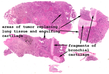 |
Very little alveolar
lung is to be found on this slide. The tumor has pretty much replaced
everything in the region of the sample. Note how the tumor surrounds
and encases the fragments of bronchial cartilage.
See this slide with the
virtual microscope.
|
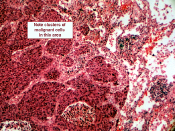 |
This slide shows a typical
squamous cell carcinoma of the lung. Mitotic figures will be common and
some "tripolar" mitoses might be present. You should be able to spot the
"intercellular bridges" (as opposed to the Madison County type) that characterize
squamous cell malignancies. You will see great variation in size of cells
and nuclei, but the basics of malignant nuclear features are all here:
nuclear/cytoplasmic ratio, angulated nuclear margins and nuclear hyperchromasia.
The type of epithelium that gave rise to this malignancy is not normally
found in the lung, where do you think it came from? |
Back
to Home
|