
General
and Systemic Histopathology, C601&C602
Slide 89: Infant lung with hyaline
membrane disease
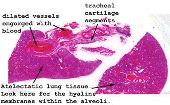 |
The lung tissue here
is quite "meaty," and may not even appear as lung to you. Fetal lung
looks much like this, although in this case there is extensive atelectasis
along with the accumulation of the alveolar proteinaceous material.
See how many aspects of pulmonary histology you can identify in this slide.
See this slide with the
virtual microscope.
|
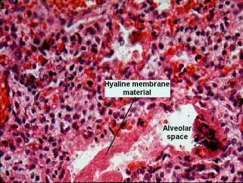 |
A higher power view of
one of the hyaline membranes. Note how the histological appearance of infant
lung is so much different from that of adult pulmonary tissue. Here you
can see how much more cellular and thick walled the alveoli are and how
cuboidal the alveolar lining epithelium appears. This cuboidal nature of
the epithelium is not a function of the disease, rather reflective of the
degree of immaturity of the lung tissue. |
Back
to Home
|