General
and Systemic Histopathology, C601&C602
Slide 113: Kidney with acute and chronic
pyelonephritis

|
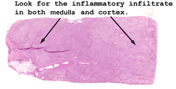
This slide shows an
extensive mixed acute and chronic inflammatory infiltrate throughout the
interstitial tissue. You will also see acute inflammatory cells in
the lumens of many of the tubules. You know this kidney must have hurt.
See this slide with the
virtual microscope. |

|
Note the marked degree
of chronic and acute inflammation in the interstitial tissue. You will see
some periglomerular fibrosis as well as marked changes in the epithelium lining
the tubules. See if you can find any "casts" still in the tubules. What would
they look like if they had been cleared in the urine?
What do you think this person felt like? Fever? Peripheral WBC count?
What organism is most likely, and how did it get here? What about antibodies
stuck to the bugs? What would this mean if observed in the urine? |
Back to Home
|