
General
and Systemic Histopathology, C601&C602
Slide 145: Kidney with clear cell carcinoma
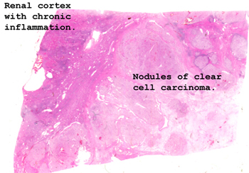 |
There should be no problem
finding the area of tumor if you simply look at the tissue on a white background.
Note the nodularity of the tumor. Also, there is quite a chronic
inflammatory infiltrate in the surrounding uninvolved renal tissue.
See this slide with the
virtual microscope. |
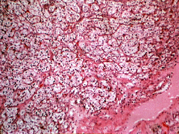 |
The entire field in this
case is composed of the carcinoma. Note how the cells reveal a clear almost
empty cytoplasm. These malignant cells contained a lipid droplet at one
time, but it was "washed out" during the processing of the tissue. This
is one of the most enigmatic tumors we know of. It can metastasize widely,
but patients may go for years with it. It often uses the existing vasculature
as a framework to grow on. You should see many mitotic figures. |
Back
to Home
|