General and Systemic Histopathology, C601&C602
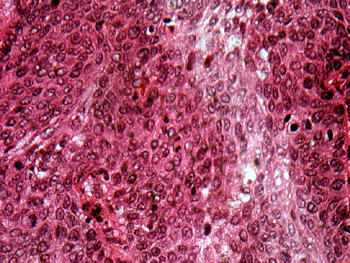
Slide 153: Squamous cell carcinoma of uterine cervix
| As with slide 61,
it's best to get the tissue oriented before going to your microscope.
The dilated nabothian glands should serve as a marker for the endocervix.
The malignancy is on the ectocervical side. Can you identify areas of
invasion? See this slide with the virtual microscope. |
|

|
In this case, the cancer is not only in the mucosa, but can be found in the superficial lymphatics as well. Note the marked degree of nuclear anaplasia in the epithelial cells that are very close to the actual surface of the mucosa. There is no maturation at all. You should see numerous mitotic figures. There will be some inflammation which may obscure the process in some areas. Scan this with low power so you know where to concentrate your efforts. |
Reproductive Diseases Lab Page
| Previous Slide
| Next Slide
| Table of Contents
Mark
Braun, MD braunm@indiana.edu
Copyright 1998,
the Trustees of Indiana University