
General
and Systemic Histopathology, C601&C602
Slide 61: Cervical dysplasia
|
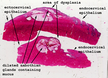
This can be a confusing
slide if you don't get oriented first. In the scan at the left you can
see both the endocervical and ectocervical regions. The big dilated
cystic, mucin filled glands in the middle are glands of Naboth. We see
these either in the endocervix or at the endocervical-ectocervical junction.
Use them as a landmark to tell where you are. If you see squamous epithelium
over these glands, it almost always represents squamous metaplasia.
In this case, there's dysplasia as well.
See this slide with the
virtual microscope. |
|
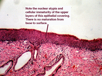
This picture pretty
well says it all. This condition is viral in nature and some of the viral
serotypes are known to be associated with the development of cancer. You
will see immature basilar cells carried far up into the epithelial covering.
There are many mitoses as well as generalized atypia of the nuclei. These
change can look pretty disturbing on a PAP stain. |
Back to Home
|