
General
and Systemic Histopathology, C601&C602
Slide 17: Skin with leishmaniasis.
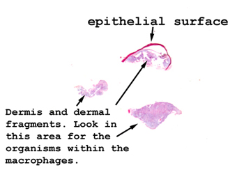 |
This specimen consists
of little fragments of skin and it's difficult to get oriented. You want
to be looking in the dermis for histiocytes containing the organisms.
There is a lot of necrosis along with the inflammatory infiltrate.
See this slide with the
virtual microscope. |
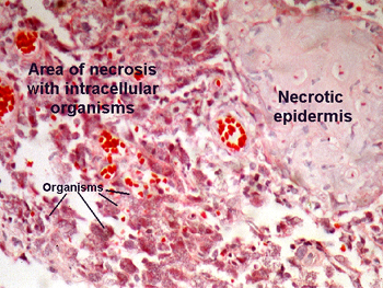 |
This slide is not H&E
stained. We used a stain to highlight the organisms. In some slides there
is an ulcer with granulation tissue in the base. Look in the dermis and
you will see an infiltrate composed largely of mononuclear cells. The organisms
are in the monocytes, and appear as small dots with a cleared area or "halo"
around them. They are quite small. Some may appear in the tissue, but I
think this reflects rupture of the cells, possibly even as a tissue processing
artifact. This slide is to further your education, and I'll tell you right
now I don't have a sample of this in my quiz slide collection. |
Back
to Home
|