
General
and Systemic Histopathology, C601&C602
Slide 52: Skin with Intradermal Nevus
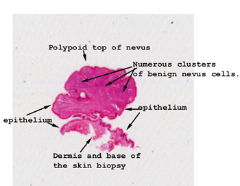 |
This picture is a nice
cross section of a piece of skin with a polypoid shaped nevus extending
from the skin surface. Not all nevi acquire this polypoid configuration
but a fair number do. Your section may not include the stalk, sorry.
See this slide with the
virtual microscope. |
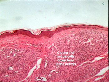 |
Note the clusters of
benign nevus cells in the dermis. There is maturation "from surface to
base," and by this we mean the nevus cells look more and more mature as
you scan from the epidermal covering to the deeper dermis. You will see
no mitotic figures and there is no cytoatypia of the nevus cells. If there
had been nevus cells in the epidermis what would have the lesion been called?
No the answer is not melanoma. |
Back
to Home
|