
General
and Systemic Histopathology, C601&C602
Slide 121: Chronic Myositis
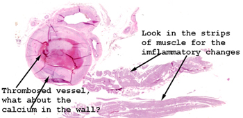 |
Obviously there are several
pieces of tissue on this slide. Look in the strip of muscle.
You are looking for inflammatory cells in between the individual muscle
cells as well as disruption of the actin and myosin within the cells.
See this slide with the
virtual microscope. |
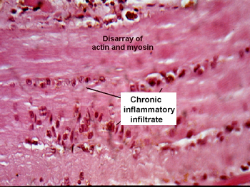 |
You will see many "ratty"
looking and fragmented bundles of muscle cells, as well as disrupted actin
and myosin within a few of the individual myocytes. There should be no
trouble finding the chronic inflammatory infiltrate located between the
muscle cells. On low power you should be able to appreciate the great variation
in muscle cell diameter. What types of conditions would bring about this
change? What serum enzymes would you expect to be elevated in a patient
with such a condition? |
Back
to Home
|