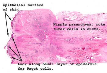
Here we have the condition
of malignant duct cells growing up the major excretory ducts of the breast
and out into the epithelial covering of the skin. You will actually
see clusters of malignant cells in the epithelium itself. Look down
in the breast to confirm the malignancy first. Sometimes this can be
confused microscopically with an early amelanotic melanoma. Grossly,
this lesion is red and crusted and looks like a little focus of irritation
on the nipple or areola.
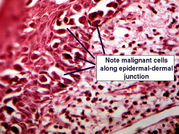
Here you see a higher
power view of the clusters of the malignant ductal epithelial cells which
have actually migrated up and out of the major breast ducts to proliferate
within the epithelial layer of the skin. These cells may look a bit like non-pigmented
malignant melanocytes, but they are indeed from the breast ducts. The cells
you see here are not within dermal lymphatics, but rather the actual epithelial
surface of the breast itself.