
General
and Systemic Histopathology, C601&C602
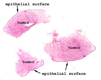
Slide 33: Skin with metastatic breast cancer
|
This situation is different
from what we looked at in slide 31. Here we actually see little dermal
implants of metastatic breast cancer, not epithelial spread. These metastases
can be from anywhere, unlike the situation of Paget's disease depicted in
slide 31, where the spread is by direct continuity to the overlying nipple
skin.
See this slide with the
virtual microscope. |
|
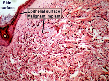
Note the difference
between this slide and #31. The malignant cells are in the dermis and represent
a distant metastases, not a direct "creeping type" spread from the breast
ducts below the epidermis. This pattern is more typical of metastatic breast
cancer. Take a look for the single file arrangement and "pseudoglandular"
organization of this tumor. This pattern is highly characteristic which makes
it possible to make a very good guess as to the primary when presented only
with the metastatic tumor. |
Back to Home
|