
General
and Systemic Histopathology, C601&C602
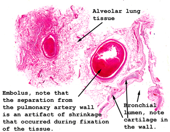
Slide 14: Lung with pulmonary embolus
|
The picture pretty
much says it all. You should have no trouble finding the clots in the
pulmonary vasculature.
See this slide with the
virtual microscope. |
|
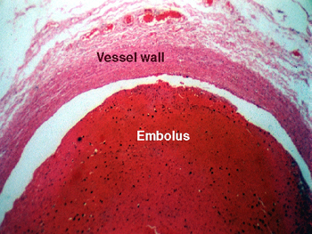
This slide shows a
pulmonary embolus lodged in a major pulmonary artery. An embolus is a blood
clot that formed somewhere else, broke free, traveled through the vascular
system and lodged in the pulmonary vasculature. Do you know
the difference between a thrombus and embolus
? Many things can become an "embolus," the term is not specific: bullets,
bone chips, amniotic fluid, even air. This slide shows a fairly typical artifact
of formalin fixed tissue. At the time of death, the blood clot filled the
vessel, the area of "clearing" between the vessel wall and the clot that
we see now represents shrinkage during processing of the tissue. |
Back to Home
|