These pictures pretty
much say it all. Look how markedly narrowed the lumen is in some of
these serial sections. You can easily see the calcium deposits in some
of the plaques. Obviously it wouldn't take much to fully occlude the
lumen in several of these sections.
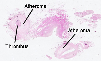
This picture is of
only a small portion of the vessel wall. Note the calcium in the atherosclerotic
plaque (blue crystals), and the "cholesterol slits." The plaque has a complex
structure and starts out as an intimal lesion. Other conditions can have calcium
deposits in the walls of vessels, but the deposits of an atheroma occur in
the subendothelium. If thrombus should form on the surface of the plaque,
the vessel will become completely occluded. Sometimes the material of the
plaque can "embolize" i.e. break free and head down stream, causing small
vessel arterial occlusion at a location distant from the plaque itself.