
General
and Systemic Histopathology, C601&C602
Slide 190: Kaposi's Sarcoma of the
Skin
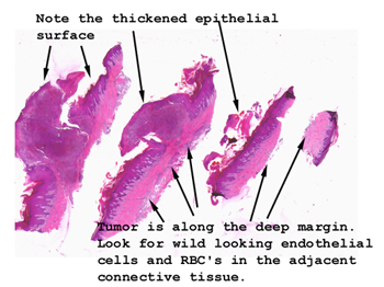 |
Here we have multiple
serial sections of the skin biopsy. Once you've scanned the entire
slide, look in the dermis for the characteristic malignant endothelial
cells.
See this slide with the
virtual microscope. |
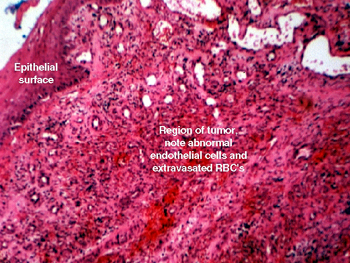 |
Yes, this biopsy is from
an AIDS patient. Note the extravasated RBC's in the surrounding connective
tissue and the bizarre and very disturbed looking endothelial cells that
comprise this lesion. You will see many very abnormal "fibroblast" looking
cells, but these are really bizarre endothelial cells. You will also see
some dark brown or black pigment in the background and possibly in some
of the histiocytes. This pigment is actually iron from the red blood cells
that have previously broken down. An iron stain, such as a Prussian Blue,
would really highlight this feature. |
Back
to Home
|