
General
and Systemic Histopathology, C601&C602
Slide 58: Lung with abscess
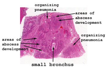 |
There is such consolidation
in this section of lung, that it is hardly recognizable for what it is.
Just looking at the piece of tissue on the slide, you'll the diffuse infiltrate
as well as the darker areas represent the complete breakdown of the pulmonary
tissue. Start by looking at the edge of the tissue to see if you
can find any "normal" lung to help you get oriented.
See this slide with the
virtual microscope. |
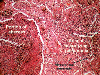 |
This condition could
be due to any number of bacterial organisms, or even a mixture of bugs,
but it happens to be staphylococcus. The general alveolar outlines will
be hard to find in the areas of necrosis. It will help to go to the edge
of the tissue to get your bearings before going to the area of the lesion.
You will need to be pretty familiar with normal lung architecture to see
anything in the background. Much of the lung parenchyma has been destroyed
by the digestive enzymes of the bugs. You will see many acute inflammatory
cells along with the amorphous digested debris. Clearly, once the lung
tissue has been destroyed and the abscess formed, that lung tissue is gone
for good. |
Back
to Home
|