
General
and Systemic Histopathology, C601&C602
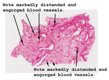
Slide 74: Lung with passive congestion
|
The congested vessels
literally leap out of the tissue on this slide. Look not only at the vessels,
but also the frothy material that has collected within the alveolar spaces.
What's going on here?
See this slide with the
virtual microscope. |
|
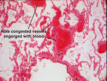
The changes here are
a little subtle. The blood vessels are distended and "over filled" with blood.
They have became dilated, in this case, because of the back up of blood
in the pulmonary circulation due to an abruptly failing left ventricle. This
person had a myocardial infarction, and experienced sudden failure of the
pump. You may see evidence of pulmonary edema in the alveolar air spaces.
The edema fluid will appear as a faint pink stained material in the background
of the air spaces. It represents the extravasation of fluid through the vessel
wall as a result of the increased lumenal pressure. |
Back to Home
|