
General
and Systemic Histopathology, C601&C602
Slide 168: Skin with Amyloidosis, H&E
Stain
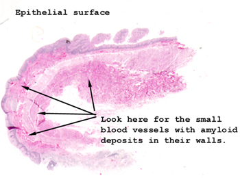 |
You want to look at the
vessels in the dermis. The amyloid deposits will be in the walls.
See this slide with the
virtual microscope.
|
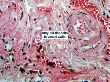 |
Here you can see a slight
enhancement of the eosinophilic staining of the vessel walls, but not nearly
to the extent when the diagnostic congo red stain is used. Concentrate
in the walls of the vessels in the dermis and compare your observations
with slide 169. |
Back
to Home
|