 Simple
columnar (Fig. 4-13) Simple
columnar (Fig. 4-13)
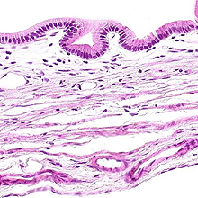
- Simple columnar epithelia are
specialized for absorption.
- This type of epithelium lines
the gall bladder (slide 3).
- The projecting villi in the
small intestine (slide 4).
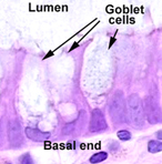
- The apical ends of these
epithelial cells are covered by microvilli to provide a larger
surface for absorption.
Examine both the LM and EM
pictures
of microvilli in Fig. 4-8.
- On
slide 4 the
glycocalyx on the
microvilli is stained "magenta" by the PAS reaction,
demonstrating the microvilli colle
 ctively
as the brush border or striated border. ctively
as the brush border or striated border.
- On
slide 21, stained with trichrome, study the brush border again and examine the
terminal
web, the network of actin filaments in the cytoplasm just below
the microvilli.
- Draw several columnar
epithelial cells, showing the nuclei, the apical ends,
microvilli, and glycocalyx.
- Some cells of the simple
columnar epithelium on slide 4, called
goblet cells, are
specialized for secretion rather than absorption.
- PAS stains the mucin
granules which fill the cytoplasm, demonstrating the overall
size and shape of these cells. See also Fig. 4-17
Here are some interesting facts about epithelial
cells. |