Simple cuboidal (Fig. 4-12)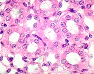
- The sample of kidney on
slide 14
shows many examples of simple epithelia, from
very thin simple squamous to rather high cuboidal.
- In the image to the right,
we're looking at renal tubules lined with a single layer of
simple cuboidal epithelium.
- The bright blue color in the
Masson trichrome stain used here stains collagen and helps
demonstrate the basement membranes of the epithelia.
- What are the
structural differences between the apical and the basal
“sides” of cuboidal epithelial cells?
Now for simple
columnar epithelium. |