 Simple
squamous (Fig. 4-11) Simple
squamous (Fig. 4-11)
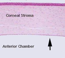
- First examine the eye (slide
78), where simple squamous epithelia can be found as the inner
lining of the cornea, the corneal endothelium. (Fig. 23-4 will help you find this layer of the eye if necessary.)
- What is located in the
thickest part of a simple squamous cell?
-
 Then
examine some "loose” or areolar connective tissue (slide 45)
where blood vessels of all sizes can be found. The lining of the
smaller vessels (vascular endothelium) provides a good example
of this epithelial type, shown in Fig. 4-1. Then
examine some "loose” or areolar connective tissue (slide 45)
where blood vessels of all sizes can be found. The lining of the
smaller vessels (vascular endothelium) provides a good example
of this epithelial type, shown in Fig. 4-1.
- To the right is an image
of a gallbladder with small vessels in the lamina propria.
- Why doesn’t every
region of the vascular endothelial lining show cell nuclei?
Now for simple
cuboidal epithelium. |