 Lung
-- organ in which bronchi become highly branched and in which
respiratory exchange occurs (Fig. 17-6). Lung
-- organ in which bronchi become highly branched and in which
respiratory exchange occurs (Fig. 17-6).
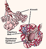
Study the functional organization of lung tissue as shown in Fig.
17-6 and on the drawings here, noting
especially the extra- and intrapulmonary airways and the
intrapulmonary blood circulation around the alveoli.
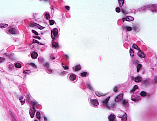
Examine an H&E-stained section of the
lung (slide 97).
- Locate an uncut edge showing visceral pleura
covered by a simple squamous epithelium, the mesothelium (Fig.
17-18).
- Deeper in the lung, identify the small
bronchioles (Fig.
17-9), which lack any cartilage framework and usually run parallel
to small arteries or arterioles. (This slide does not include larger
branches of bronchi, Figs. 17-7 and 17-8.)
- Note the bronchioles’ respiratory epithelium
and the thin layer smooth muscle in their lamina propria layer.
Compare and contrast the
structure of the bronchioles in this slide with that of the trachea.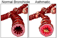
Clinical note: Asthma involves
hyperirritability of the respiratory passages, expressed as
contraction of the bronchial smooth muscle, edema of the mucosa, and
increased mucus secretion. It may be transient, following an upper
respiratory tract infection, but more commonly the sensitization has
an immunological basis and the symptoms are episodic. Re-exposure to
an airborne antigen such as pollen causes release of histamine from
mast cells and eosinophils, precipitating immediate
bronchioconstriction and labored breathing. Various drugs are
helpful in minimizing the severity of the attacks.
Elastic fibers
in the lung. |