|
 Clinical
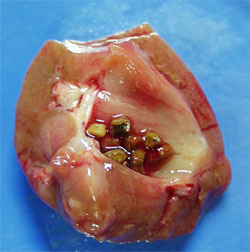
note: Kidney stones (nephrolithiasis) are concretions of calcium
salts and uric acid that can form in the renal pelvis when urine
contains high concentrations of substances such as calcium and uric
acid. Small stones may pass down the ureters (with considerable
pain) and larger stones can be destroyed by focused sound waves in a
procedure called lithotripsy. (Photo courtesy of Dr. C.F. Verkoelen) Clinical
note: Kidney stones (nephrolithiasis) are concretions of calcium
salts and uric acid that can form in the renal pelvis when urine
contains high concentrations of substances such as calcium and uric
acid. Small stones may pass down the ureters (with considerable
pain) and larger stones can be destroyed by focused sound waves in a
procedure called lithotripsy. (Photo courtesy of Dr. C.F. Verkoelen)
Trace the vasculature and
microvasculature through the kidney in the diagram (Fig. 19-3) and
indicate which vascular components are associated with specific
regions and nephron components.
Examine the structures in the cortex and
medulla in greater detail using the section of human kidney on
slides 14 and
144.
- Study the diagram of a renal
corpuscle (Fig. 19-5) and examine them on the slides to identify
the simple squamous epithelium of Bowman's capsule, glomeruli,
the large, pale podocytes (Fig. 19-14b).
- Study the electron micrographs
of glomeruli (Figs. 19-5 and 19-6), identifying
- Podocytes,
- Capillary endothelial cells,
- The intervening basement
membrane, and
- Bowman's space.
- Note especially the
organization of the podocyte foot processes and the
endothelial cells fenestrations at the basement membrane.
- On the slide look at many
examples of glomeruli to find one cut to show one or both of
the afferent or efferent arterioles (Fig. 19-5).
Describe the structural
features which produce the process of renal filtration.
What is significance of
having afferent and efferent arterioles in this process?
What drains Bowman’s
space? Next
let's consider kidney infections and
look at examples of he nephron. |