|
 Kidney
-- a bean-shaped organ with renal arteries and veins entering with
the ureter at the hilum. Kidney
-- a bean-shaped organ with renal arteries and veins entering with
the ureter at the hilum. Study
the diagrams showing the overall organization of the kidney and the
note the arrangement of the components in nephrons (Figs.
19-1, 19-2
and the next page) and examine the preserved-mounted, hemisected
specimens of kidney, noting all the macroscopic features indicated
in the diagrams. Examine a
Masson trichrome-stained section of the kidney (slide 14)
- Note the thin fibrous capsule
and the fibrous/fatty support tissue at the hilum area
surrounding the renal pelvis (Fig. 19-1). The orientation of
this section does not clearly show the kidney's internal
organization.
 For this examine
slide 111, a section of rat
kidney which unlike the human kidney is unilobular, i.e.,
consists of one large lobe with ducts converging in the
direction of the hilum. For this examine
slide 111, a section of rat
kidney which unlike the human kidney is unilobular, i.e.,
consists of one large lobe with ducts converging in the
direction of the hilum.
- With low power, identify the
cortex and the medulla (Fig. 19-2).
- Near the corticomedullary
junction can be several sets of arcuate arteries and veins (Fig.
19-3) cut transversely. (The veins still contain blood.) Still
with low power, identify in the cortex renal corpuscles (Fig.
19-4) and then the medullary rays converging on the renal
papilla and calyx (Fig. 19-1).
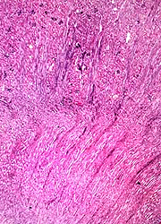
- Note that the largest collecting
ducts (the ducts of Bellini) converge to form the renal papilla,
and that this is surrounded by the calyx composed of
transitional or urinary epithelium (Fig. 19-2).
What features principally
distinguish renal cortex from medulla?
What constitutes a “renal lobe?”
Are the calices considered
part of the medulla?
Kidney stones
and the renal cortex. |