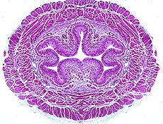
 Esophagus
-- conducts food from oral cavity to stomach. Start by reviewing the
general plan of the gastrointestinal tract (Fig. 15-2), noting
especially the four major layers which are clearly seen in the
esophagus: Esophagus
-- conducts food from oral cavity to stomach. Start by reviewing the
general plan of the gastrointestinal tract (Fig. 15-2), noting
especially the four major layers which are clearly seen in the
esophagus:
- Mucosa
- The epithelial lining of various
types
- Lamina propria of connective
tissue
- Thin muscularis mucosae of
smooth muscle
- Submucosa containing blood
vessels, lymphatics, and nerves
- Muscularis: two thick layers of
smooth muscle for peristalsis
- Adventitia or serosa: outer
connective tissue covering
Examine the preserved-mounted
specimens of the various regions of the digestive tract, noting the
macroscopic features (stomach rugae, plicae circulares, etc.) for
correlation with the slides you will also study.
Examine a transverse section of the
esophagus (slide 66 or
43). Identify the various
layers and sublayers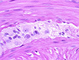 indicated in the general plan (Fig. 15-14)
- Note the epithelial type of the
lining,
- The presence of small mucous
glands
- Any lymphoid nodules present, and
- Autonomic ganglia and nerves of
the myenteric plexus (Fig. 15-35b).
What would the muscularis in the
upper region of the esophagus show differently?
Let's consider the
muscular layers in more detail. |