 For
the study of the major salivary glands, examine sections of the
parotid gland (slide 132) and the submandibular gland (slide
11 and
72). For
the study of the major salivary glands, examine sections of the
parotid gland (slide 132) and the submandibular gland (slide
11 and
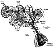
72).Such a gland is shown
schematically in Fig. 16-2 and in this diagram, which also shows the
comparison with the pancreas which is histologically very similar to
the major salivary glands. Click the image for an
expanded view.
-
Acini of the parotid gland (Fig.
16-3) are completely serous.
- Submandibular gland
(Fig. 16-6) are mixed serous and mucous.
- The PAS -alcian blue stain of
slide 11 stains mucus well and shows distinct difference between
the types of secretory cells.
- Compare serous and mucous
secretory cells in these slides
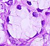 . .
- On slides
72 and
11, identify the
mucous and serous cells and look for serous demilunes (Fig.
16-6).
What do serous and mucous cells
respectively contribute to saliva?
What could be the functional
significance of serous demilunes?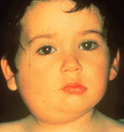
Clinical note: The childhood
disease called mumps is a viral infection of the salivary glands,
almost always the parotid glands, causing swelling and tenderness
for a week or so. The disease is
usually self-limiting but the virus can spread to other organs,
including the inner ear where it can lead to
deafness.
Look for striated ducts (Fig. 16-5)
and larger excretory ducts in both slides
11 and
132.
What unusual types of epithelia
are found in the ducts of salivary glands?
What accounts for the basal
striations in the epithelial cells of the striated ducts?
Now for the esophagus. |